PET Medical Examination
PET Medical Examination
General overview of operations
Please apply to Wellness Medical Information Center when you reserve it.
Depending on the medical care institution, the discount might not apply.
Reservations/inquiries:
03-6706-7384
User Subsidies
The following subsidies are available to insured individuals aged 35 years or over when they undergo a PET medical examination.
(Assistance is not available to insured individuals aged 35 years or under or to dependents.)
| Medical examination including neurological examination | 15,000 yen
|
|---|
Charges
For details on medical care institution fees, please inquire with the Wellness Medical Information Center.
Appointments and inquiries: 03-6706-7384
- * Please tell them that you are a member of the Bosch Health Insurance Society.
WELLNESS MEDICAL INFORMATION CENTER
Brain Dock (Medical Examination of Brain) Expense Subsidy Bill
General definition of cancer
The human body is made up of approximately 60 trillion individual cells, a number that is almost unimaginable. These cells are normally regulated by our genes such that at any one time we have the appropriate total number of cells in our bodies. However, in some cases, for some reason cells suddenly appear that disrupt this normal genetic behavior and continue to multiply in an uncontrollable manner. These cells are known as "cancer cells."
Cancer's frightening stealth
Until recently, cancer was generally regarded as an incurable illness. However, testing and treatment technologies have advanced to the stage at which this illness is on the verge of becoming surmountable, as long as it is detected early. However, because in its early stages cancer exhibits few subjective symptoms (such as pain or discomfort) and because cancers take a long time to develop to the point at which they are visible to the naked eye, in a considerable number of cases the disease is already difficult to treat when first detected.
Methods of testing for cancer
PET NET JAPAN-affiliated PET facilities offer PET scans as well as numerous other types of tests. The combination of various tests with a PET scan offers patients more accurate results.
| CT scan | In a CT scan, X-rays are used to detect irregularities in the shape of the patient's internal organs. Because X-rays are irradiated onto the body at all angles within a full 360 degrees, the CT scan produces images that show cross-sections of the body, a result difficult to achieve in normal X-ray testing. During cancer examinations, CT scans are mainly used to detect lesions in the bronchi, lungs, or other areas of the chest, or in the liver, kidneys, or other areas of the abdomen. |
|---|---|
| Ultrasound | In an ultrasound (sometimes referred to as an "echo test"), ultrasonic waves are directed at the body from an external source. The "echo" waves reflected from within the body are used to create an image that is then used to check the condition of internal organs and tissues. There is absolutely no pain or radiation exposure, rendering ultrasound testing ideal when assessing a wide range of areas, such as thyroid glands, mammary glands, the abdomen, and the pelvis. |
| MRI scan | MRI scans use powerful magnetic forces and magnetic fields to assess the condition of internal organs. In addition to producing cross-sectional images from any angle without shifting the patient's body, MRI scans can also produce detailed images of specific targets such as blood-vessel shapes. MRI scans are particularly effective in examining the pelvis and head, and are also useful in detecting such cancers as uterine cancer, ovarian cancer, and prostate cancer, and in diagnosing conditions such as brain tumors and brain infarctions. |
| Biochemical marker test (tumor markers) |
Tumor marker tests are tests that determine the presence and type of cancer by taking blood, urine, or stool samples and measuring the levels of particular substances (glycoprotein, hormones, enzymes, etc.) produced when cancer cells are present, or measuring substances produced by normal cells when they react to the presence of cancer cells. There are many varieties of marker tests; combining different types enables testing for a variety of cancers at the same time. |
CT scan
Ultrasound
MRI scan
Biochemical marker test
(tumor markers)
Cancer death rates
The leading cause of death among Japanese
Cancer has been the leading cause of death since 1981.
According to the Ministry of Health, Labour and Welfare's demographic statistics, around 30 percent of Japanese people currently die of cancer, which is over 300,000 deaths per year.
Nowadays, one out of every two Japanese people is said to get cancer, so cancer is not simply someone else's problem.
Anyone could get cancer, so everyone has to be careful about it.
The importance of early detection
Above all else, the key to overcoming cancer is early detection
Cancer cells are extremely gluttonous, and weaken the body by stealing the nutrients that normal cells need in order to grow. They gradually infiltrate surrounding normal cells and spread to other parts of the body via blood vessels and lymphatic vessels, causing the lesions to spread. In order to overcome cancer, it is essential that it be detected while it still remains in the organ in which it first appeared. In other words, the key to winning the battle against cancer is the speed with which it is detected.
What is a PET scan
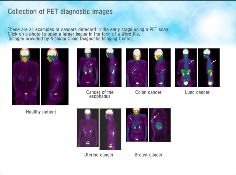
PET is a state-of-the-art diagnostic imaging technique used for the early detection of cancer.
PET is short for positron emission tomography, a state-of-the-art technique of diagnostic imaging. While traditional methods diagnose tumors based on the shape of the organs, PET scans check the metabolism and function of cells, thereby enabling more definitive diagnosis of early-stage cancer.
PET scans are also effective in determining whether a tumor is benign or malignant, and in confirming whether cancer has spread or recurred.
The first stage of a PET scan involves injecting a substance (FDG) very similar to glucose into the patient's body. One characteristic of this substance is its attraction to cancer cells, and therefore it concentrates near cancer cells. Generating a corresponding image using a PET camera enables determination of the presence or absence of cancer. Also, by looking at the extent of FDG concentration, it is possible to determine whether the tumor is benign or malignant, and whether it has spread to the lymph nodes or recurred.
How a PET scan works
PET scans make it possible to examine the entire body in one go without pain or discomfort.
With the exception of the initial injection of FDG, the patient experiences no pain or discomfort when undergoing a PET scan. Moreover, it is possible to screen the entire body (from below the eyebrows to the groin) during a single scan. In addition, because patients can be scanned while clothed (in a screening gown), women in particular feel more reassured when undergoing a PET scan.
The PET scan process
Collection of PET diagnostic images